
Radiology imaging and pregnancy – what you need to know
Radiology is essential in the diagnosis of both acute and chronic illnesses. Since pregnancy is a special condition, issues on the safety of medical imaging often arise. However, to monitor the baby's progress or diagnose a certain disease, medical imaging is needed. Various safe methods of imaging can be used to image the mother or the fetus.
Radiological services can be given in public hospitals or private facilities. Mermaid Beach radiology gold coast offers world-class medical imaging services. The facility offers quality and personalized medical imaging.
Imaging Techniques
Radiological imaging comprises various techniques.
-
Ultrasonography
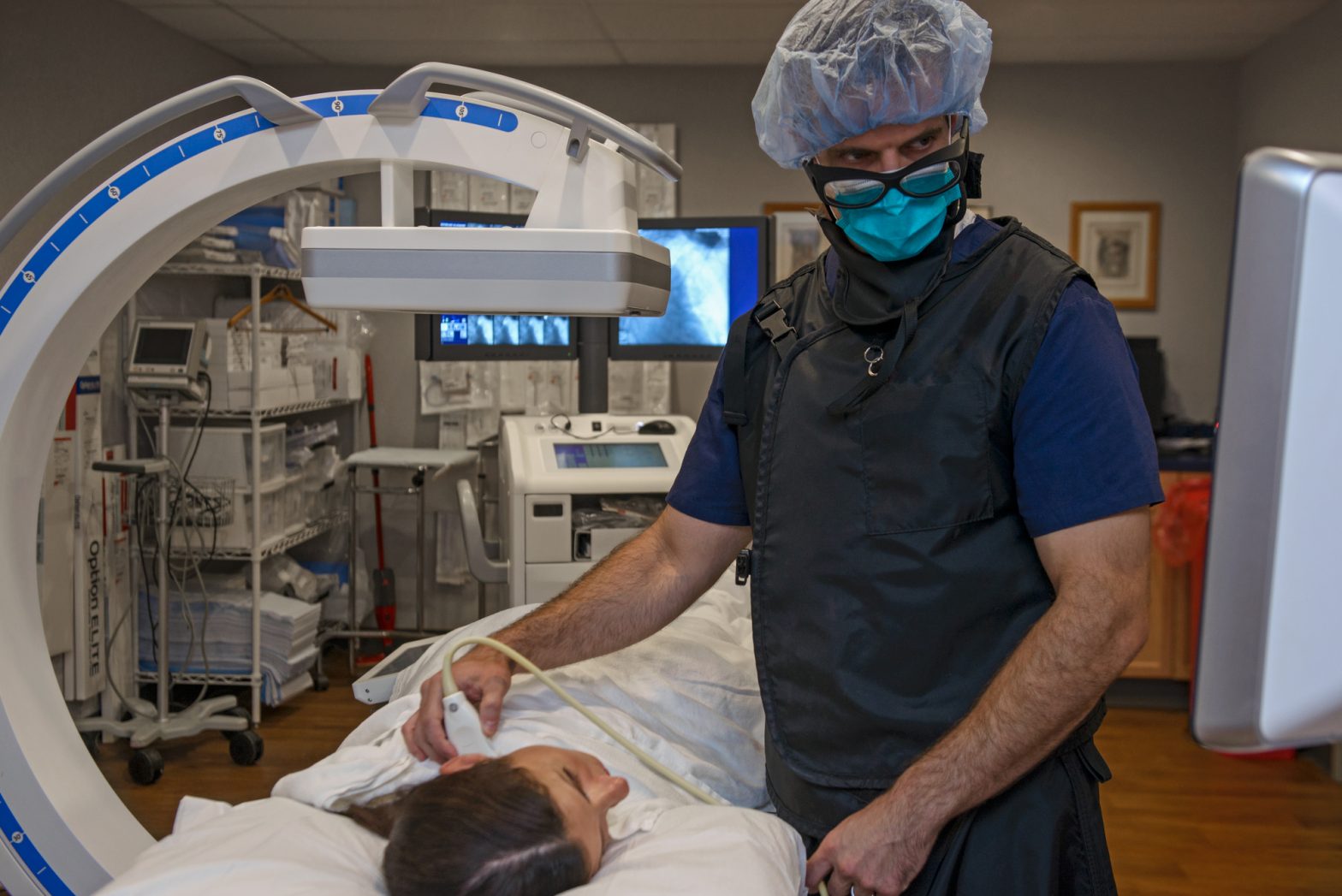
Ultrasound imaging is among the safest radiological procedures. The technique uses sound waves of high frequency to image the internal organs of the body. Sound waves have very low frequencies, therefore non-ionizing.
Ultrasound imaging has a paramount role to play during pregnancy. During the procedure, a transducer is moved over the belly. It captures images that are then displayed on a screen. In transvaginal ultrasonography, the transducer is inserted through the vagina.
The uses of ultrasound in pregnancy include:
- Confirmation of pregnancy
- Checking for multiple or ectopic pregnancy
- Examining the uterus and the placenta
- Diagnosis of fetal abnormalities
- Monitoring the growth of the fetus
- Guiding amniocentesis
- Determining the sex of the baby during the third or second trimester

-
Magnetic Resonance Imaging
For detailed 3-D images, a magnetic resonance imaging (MRI) scan is recommended. The technique uses magnetic fields alongside radio waves to image the body organs. It is, therefore, non-ionizing. The technique efficiently detects abnormalities such as tumors, aneurysms, stroke, and bleeding. MRI technique is considered safe for imaging during pregnancy.
MRI is commonly used during pregnancy to diagnose diseases affecting the mother. Besides, it is used during the examination of complications associated with pregnancy. It is preferred since it gives detailed information and it is relatively safe. On the other hand, ultrasound is considered the method of choice for fetal imaging.

-
Computerized Tomography and X-rays
Computerized tomography (CT) scan uses x-rays. Both CT and x-ray scans are rarely used for imaging during pregnancy. However, if it is necessary that an x-ray or CT imaging has to be used, the benefits and risks must be carefully weighed. In such a case, these low doses of ionizing radiation are used.
X-rays are ionizing radiations. High doses can result in:
- Fetal abnormalities
- Fetal death
- Growth restrictions
- Brain and intellectual dysfunction
- Congenital anomalies

-
Nuclear Imaging
Nuclear medicine imaging uses radioisotopes to assess the physiological function of a given organ. Radioisotopes are unstable elements that, on decaying, emit radiation. The emitted radiation is quantified for diagnosis. The techniques may be employed for imaging the expectant mother's body organs.
Safety concerns revolve around the type of radioisotope used. Low-dose technetium 99m is considered relatively safe during pregnancy. Iodine 131, which is commonly used to image the thyroid gland, is unsafe during pregnancy. Iodine 131 crosses the placenta and can affect the fetus' thyroid gland.
Radiology is not contraindicated in pregnancy. Thus, it can be used if necessary. Magnetic resonance imaging and ultrasonography techniques are preferred for imaging during pregnancy. The use of techniques utilizing ionizing radiations for imaging should be avoided during pregnancy. However, when necessary, these techniques are used with caution.